Introduction
Light microscopy, or optical microscopy, is a technique that uses visible light and lenses to image microscopic objects that cannot be seen with the naked eye. It has revolutionized our understanding of the microscopic world and has allowed us to view the intricate structures of cells, tissues, and organisms. Among the significant discoveries made possible by light microscopy is the observation of the first cell, a breakthrough that has had a vast impact on science. In this article, we will trace the history of the discovery of the first cell viewed through a light microscope, highlight its significance, and discuss its future in scientific research.
Tracing History: The First Organism Seen Through A Light Microscope
The invention of the light microscope is a crucial milestone in the scientific pursuit of understanding the world. The first light microscope was a simple instrument that used a single convex lens to magnify objects. It was invented in the late 16th century by two Dutch spectacle makers, Zaccharias Jansen and his father Hans Jansen. The first light microscope had a magnification power of 10x, and it was used to study objects such as insects and other small specimens.
However, it was a century later when improvements to the microscope were made, including the use of an objective lens and an eyepiece. These improvements enabled a higher magnification power and a clearer image. These advancements led to the discovery of the first organism seen through a light microscope.
Uncovering the Discovery: How Antonie van Leeuwenhoek Changed The World With A Simple Microscope
The credit for discovering the first cell viewed through a light microscope goes to Dutch scientist Antonie van Leeuwenhoek. Van Leeuwenhoek was a draper who had no formal scientific training but had a knack for crafting lenses and grinding glass. It was through his work as a draper that he had access to fine threads, which he used to make microscopes with a magnifying power of 300x. These simple microscopes were small and handheld, but their high magnification power allowed scientists to see microscopic objects like never before.
In 1674, while examining the plaque on his teeth under a microscope, Van Leeuwenhoek discovered bacteria, protozoa, and other microorganisms. He later published his findings and sent letters to the Royal Society in London, describing the creatures he observed. It wasn’t until ten years later, in 1684, that Van Leeuwenhoek discovered the first cell when he observed a sample of cork under his microscope.
The Moment of Truth: What Did Robert Hooke See Through His Light Microscope?
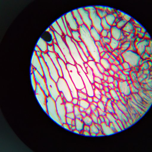
Robert Hooke, an English naturalist, had also been studying objects under a microscope around the same time as Van Leeuwenhoek. In 1665, Hooke published his findings in his book called Micrographia. In this book, he illustrated his observations of a piece of cork viewed through his microscope. Hooke described the structure he saw as a series of small compartments, which he named “cells.” It was later discovered that these compartments were the cell walls surrounding the plant’s dead cells.
The Birth of Microscopy: Lessons to Learn From The Discovery of The First Cell
The discovery of the first cell viewed through a light microscope marked the beginning of a new era in scientific exploration of the microscopic world. It revealed a new level of complexity and organization to minute living beings and their environments. It showed us how life can exist in forms beyond what our naked eyes could perceive. As we look around today, the discoveries of cells, especially in medicine and biology, could not be more precious to us.
From An Idea to A Breakthrough: The Quest for The First Cell Observed With A Light Microscope
The exploration that led to the discovery of the first cell was long and complicated. Microscopy has been critical in unlocking the mysteries of life. Over the years, scientists have made significant advancements in microscopy technology, leading to sharper images, higher magnification, and better resolution. Today, microscopy has become a standard tool in scientific laboratories and research facilities around the world. Modern techniques in microscopy include fluorescence microscopy, confocal microscopy, and scanning electron microscopy. Each has its advantages and limitations when it comes to specific applications.
Looking Back: The Five Scientists That Paved The Way To The Discovery Of The First Cell Observed With A Light Microscope
Several scientists contributed to the discovery of the first cell viewed through a light microscope. Though Antonie van Leeuwenhoek is credited with discovering the first cell, his discovery builds on the insights and information passed down by previous scientists. Among these are Galileo Galilei, who invented the compound microscope; Johannes Kepler, who built upon Galilei’s microscope design; and Robert Hooke, who first used the term “cell.”
The Basics Of Light Microscopy: How It Works, Its Advantages And Its Limitations
Light microscopy works by passing light through a specimen, which then passes through a series of lenses, forming an enlarged image of the sample on the other side. The magnified image is then recorded or visualized through an eyepiece or camera. Light microscopy is advantageous because it allows for rich color images, quick analysis, and relative ease of use. However, it has its limitations, including its resolution, meaning some structures may not be visible to the naked eye due to their small size.
Conclusion
The discovery of the first cell viewed through a light microscope has paved the way for new scientific discoveries with significant implications in biology and medicine. Microscopy remains an essential tool in scientific research, and its advancements continue to be realized daily. As technology continues to progress, the discovery of new cellular structures is likely. The future is bright for light microscopy and research alike in this field.