Introduction
Cell division is a fundamental process in living organisms. Mitosis, a type of cell division, ensures that in each newly formed cell, the genetic material is replicated and divided in an organized manner. Understanding the phases of mitosis is essential in identifying abnormal cell divisions, discovering genetic mutations, and developing new treatments for diseases, including cancer. In this article, we will examine a diagram of a mitotic phase and explore the key characteristics that make it unique from other phases of mitosis.
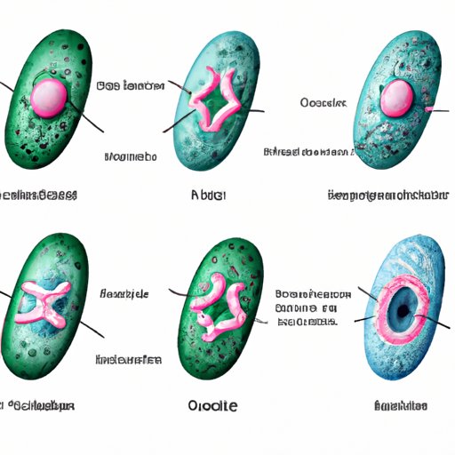
Unlocking the Secrets of Mitotic Phases: Decoding the Diagram
There are five distinct phases of mitosis: prophase, metaphase, anaphase, telophase, and cytokinesis. Each phase represents a critical step in the process where different molecular mechanisms work in concert to ensure proper cell division. Here are a few tips that can help identify the phase shown in the diagram:
- Look for key cellular structures: Each phase of mitosis is characterized by distinct cell structures that contribute to different mechanisms. For instance, during metaphase, the chromosomes align at the center of the cell, while during anaphase, the spindle fibers pull the chromosomes towards the poles.
- Assess chromosome position: Chromosomes’ position can indicate which phase is taking place. For example, in prophase, the chromosomes condense and become visible as discrete entities, but they have not yet aligned with the spindle apparatus.
Mitosis for Beginners: Understanding the Phase Shown in the Diagram
The diagram represents the metaphase phase of mitosis, which is the phase where the chromosomes align at the center of the cell. During this stage, the spindle fibers attach to the centromeres of the chromosomes, allowing the chromosomes to be divided evenly during anaphase. This stage’s completion marks a significant milestone because the chromosomes must be carefully aligned and attached to ensure that both daughter cells receive a copy of each chromosome.
Phase by Phase: A Detailed Look at Mitosis through the Diagram
Each stage of mitosis serves a unique function in ensuring proper cell division. Here is a brief look at each stage:
- Prophase: The chromatin condenses, forming discrete chromosomes. The centrosomes move to opposite sides of the cell, generating the spindle fibers that will help pull the chromosomes apart.
- Metaphase: The spindle fibers attach to the chromosomes’ protein structures called kinetochores and pull the chromosomes into alignment at the center of the cell.
- Anaphase: The sister chromatids separate and are pulled apart by the spindle fibers towards opposite poles.
- Telophase: A nuclear envelope reforms around each set of chromosomes, forming two new nuclei. The spindle fibers disassemble, and the chromosomes begin to uncoil, returning to a less condensed chromatin state.
- Cytokinesis: The cytoplasm separates into two daughter cells, each with its own nucleus, organelles, and cell membrane.
The metaphase phase is critical in ensuring that chromosomes are correctly segregated into the newly formed daughter cells. During this stage, the kinetochores, a protein structure that forms where the sister chromatids attach, are carefully aligned at the center of the cell. The spindle fibers pull the sister chromatids in opposite directions, moving them towards the poles of the cell.
Comparing and Contrasting Mitosis Phases: Analyzing the Diagram
To understand the importance of the metaphase phase, it is useful to compare it to other mitotic phases. For instance, during prophase, the chromatin condenses, and the spindle fibers form, but the chromosomes have not yet aligned. During anaphase, the sister chromatids separate, moving away from each other towards opposite sides of the cell. In contrast, the metaphase phase represents a brief period where the chromosomes are precisely aligned, and the microtubules attach to the kinetochore, ensuring that the daughter cells receive the correct chromosome numbers.
Conclusion
Identifying the different mitotic phases and understanding their specific functions is a crucial aspect of modern biology. The metaphase stage, in particular, is a critical juncture in ensuring accurate chromosome segregation. By examining the diagram and utilizing visual cues and knowledge of the molecular mechanisms that drive mitotic events, we can begin to appreciate the intricacies of cell division and its relationship to health and disease.